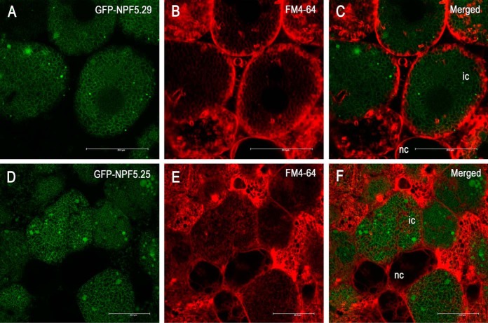
Fig. 3.
Localization of GmNPF5.29 A, B, C, and GmNPF5.25 D, E, F, to the soybean SM. Confocal images of soybean nodules expressing GFP fused to the N-terminal of GmNPF5.29, A, and GmNPF5.25, D. The SM is counterstained with, FM4–64, a lipophilic membrane stain, B and E. Overlapping GFP and FM4–64 signals are presented in the merged images, C and F. Scale bars represent 20 μm (A–F).

