Fig. 6.
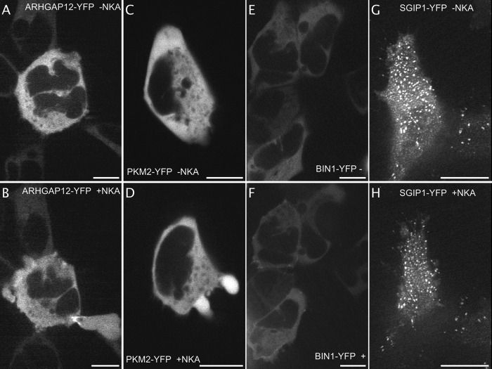
Identification of fluorescent proteins implicated in blebbing and in internalization. Images extracted from video 7 out of 9 of ARHGAP12-YFP expressing NK2R-HEK cells before (A) and 1 min after formation of the blebb (B). The protein concentrates at the opening of blebbs and their sites of resorption. Images extracted from video1 of PKM2-YFP expressing NK2R-HEK cells before (C) and few seconds after cell shrinkage (D, from video 1, 1 cell displayed out of 10). Cytoplasmic BIN1-YFP (E) transiently accumulates at plasma membrane sites around 1 min after NK2 receptors activation (F, from video 1 Part 1 out of 6 videos, 5 cells displayed out of 10). Images extracted from video1 out of 12 videos, 2 SGIP1-YFP expressing cells out of 8 before (G) and 1 min after NKA activation (H). The fluorescent protein is enriched into plasma membrane spots (probably together with clathrin) and, although cells are shrinking, NK2 receptor activation does not modify the localization of SGIP1-YFP. Scale bar 5 μm.

