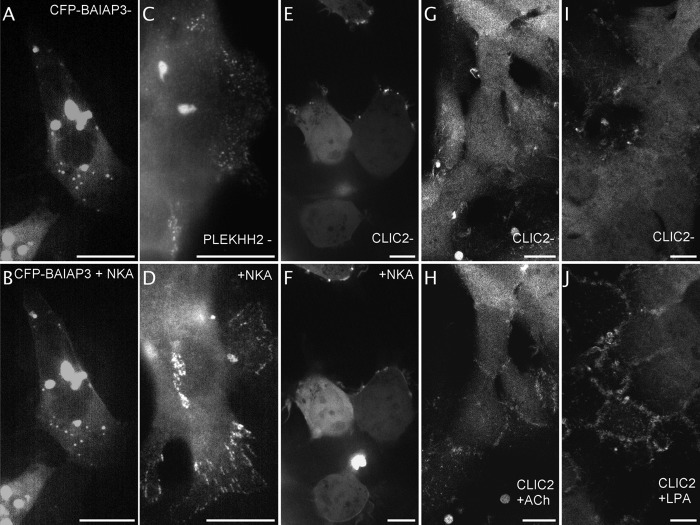
Fig. 7.
Identification of fluorescent proteins of unknown function that respond to NK2 receptors activation by a change in localization. Images extracted from videos of CFP-BAIAP3 expressing NK2R-HEK cells before (A) and within seconds after NKA activation (B, from video 1 out of 5). Representative epifluorescence images acquired close to the slide of fixed PLEKHH2-YFP expressing NK2R-HEK cells before (C) and after 3 h NKA activation (D), n = 3 independent experiments. Images extracted from videos of CLIC2-YFP expressing NK2R-HEK cells before (E) and 2 min after NKA activation (F, from video 2 out of 7 videos, 4 cells displayed out of 8). Images extracted from video1 of HEK293 cells stably expressing CLIC2-mYFP before (G) and 20 min after activation of endogenous Gq-coupled muscarinic M3 receptors with 10 μm ACh (H) n = 3 independent experiments. Images extracted from video1 of HEK293 cells stably expressing CLIC2-mYFP before (I) and 25 min after activation of endogenous G12-coupled lysophosphatidic acid receptors with 1 μm LPA (J). n = 2 independent experiments. Scale bar 5 μm.

