Fig. 1.
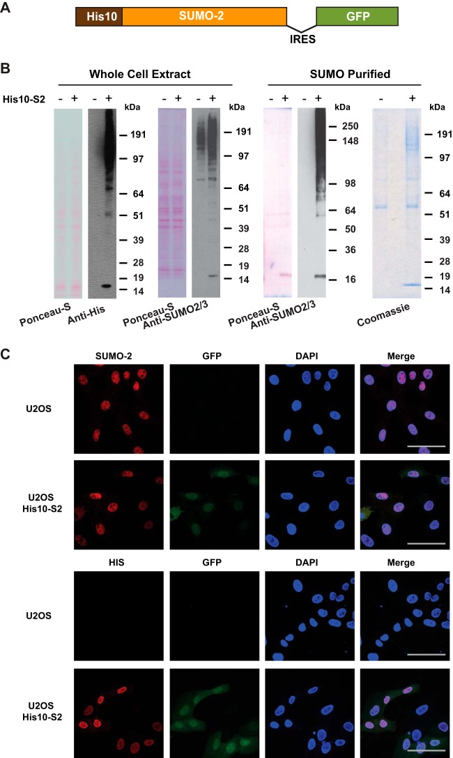
Generation and validation of U2OS cells stably expressing His10-SUMO-2. A, Schematic representation of the His10-SUMO-2-IRES-GFP construct used in this project. U2OS cells were infected with a lentivirus encoding His10-SUMO-2 (His10-S2) and GFP separated by an Internal Ribosome Entry Site (IRES) and cells stably expressing low levels of GFP were sorted by flow cytometry. B, Expression levels of SUMO-2 in U2OS cells and His10-SUMO-2 (His10-S2) expressing stable cells were compared by immunoblotting. Whole cell extracts were analyzed by immunoblotting using anti-polyHistidine and anti-SUMO-2/3 antibody to confirm the expression of SUMO-2 in U2OS cells and His10-SUMO-2 (His10-S2) expressing stable cells. Ponceau-S staining is shown as a loading control. Additionally, a His-pulldown was performed using Ni-NTA agarose beads to enrich SUMOylated proteins, and purification of His10-SUMO-2 conjugates was confirmed by immunoblotting using anti-SUMO-2/3 antibody. Ponceau-S staining and Coomassie staining were performed to confirm the purity of the final fraction. The experiment was performed in three biological replicates. C, The predominant nuclear localization of His10-SUMO-2 was visualized via confocal fluorescence microscopy after immunostaining with the indicated antibodies. DAPI staining was used to visualize the nuclei. Scale bars represent 75 μm.

