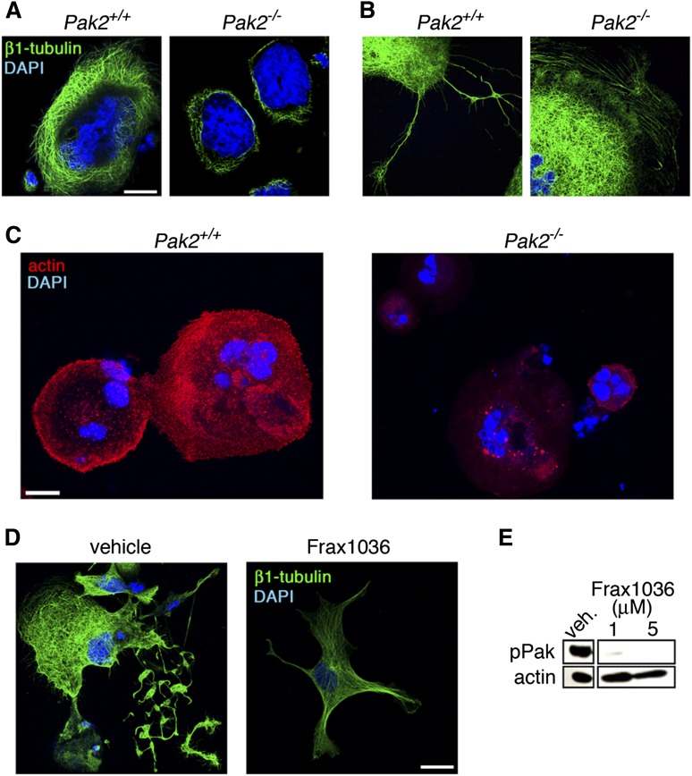
Figure 5.
Altered cytoskeleton structure in Pak2-null megakaryocytes. (A) Analysis of β1-tubulin structure and (B) proplatelet structure by fluorescence microscopy of wild-type and Pak2−/− megakaryocytes adhered to fibrinogen for 5 hours. Bone marrow treated with 500 nM 4-hydroxytamoxifen to induce Cag-Cre-ERT2 expression and delete Pak2fl/fl. Representative image of β1-tubulin (green) and DAPI nuclear (blue) staining from 3 mice per genotype. (C) Representative Alexa Fluor 594-phalloidin staining for actin (red) and DAPI (blue) analyzed by fluorescence microscopy of wild-type and Pak2−/− megakaryocytes adhered to collagen for 5 hours. (D) Fetal liver-derived megakaryocytes treated with Frax1036 for duration of culture (4 days) and stained for β1-tubulin (green) and DAPI (blue). (E) Western blot detection of serine 141 phosphorylated Pak1/2/3 (pPak) of fetal liver-derived megakaryocytes treated with Frax1036. Actin serves as a control for protein loading. Scale bar = 20 μm for all images.

