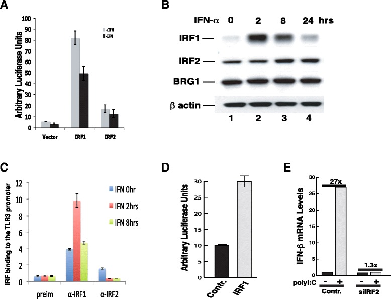
Figure 3.

The binding of IRF1 and IRF2 to the TLR3 promoter is regulated by their protein levels. A IRF1 is a strong activator whereas IRF2 is a weak activator. A luciferase reporter construct containing five GAL4 DNA binding sites was co-transfected to HeLa cells with a construct expressing either the GAL4 DNA binding domain-IRF1 fusion or the GAL4 DNA binding domain-IRF2 fusion protein for 48 hours, followed by treatment with IFN-α for 12 hours. Luciferase activity was measured as in Figure 1A. B IRF2 is expressed constitutively and IRF1 is rapidly induced by IFN-α treatment. HeLa cells were treated with IFN-α for various times as indicated above the panel. The cells were harvested and analyzed for the protein levels of IRF1, IRF2, and BRG1 using Western blotting. β actin was used as a loading control. C IRF1 and IRF2 binding to the TLR3 promoter. HeLa cells were treated with IFN-α for various times as indicated within the panel. ChIP assays were performed and analyzed using q-PCR as in Figure 2A & B. D Over-expression of IRF1 activated TLR3 promoter. pGL3-TLR3pr-luc was co-transfected with a control vector or IRF1 expression vector, pcDNA4-IRF1 into HeLa cells for 48 hours as indicated below the panel. The luciferase activity was determined as in Figure 1A. E IRF2 is required for induction of IFN-β by polyI/polyC. HeLa cells transfected with the control or siIRF2 constructs were selected in puromycin for two days. Following treatment with 20 μg/ml of polyI/polyC for 6 hours, the total RNAs were isolated and analyzed for the presence of IFN-β mRNA by real-time PCR.
