
Figure 4.
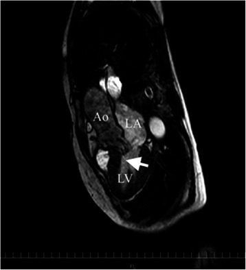
Cardiac MRI obtained 4 days after surgery. It revealed systolic anterior motion (arrow). There were no abnormalities in global shape, size, and systolic function of the left ventricle, except for some protrusion of the basal interventricular septum towards the left ventricular outflow tract. LA = left atrium; LV = left ventricle; Ao = aorta.