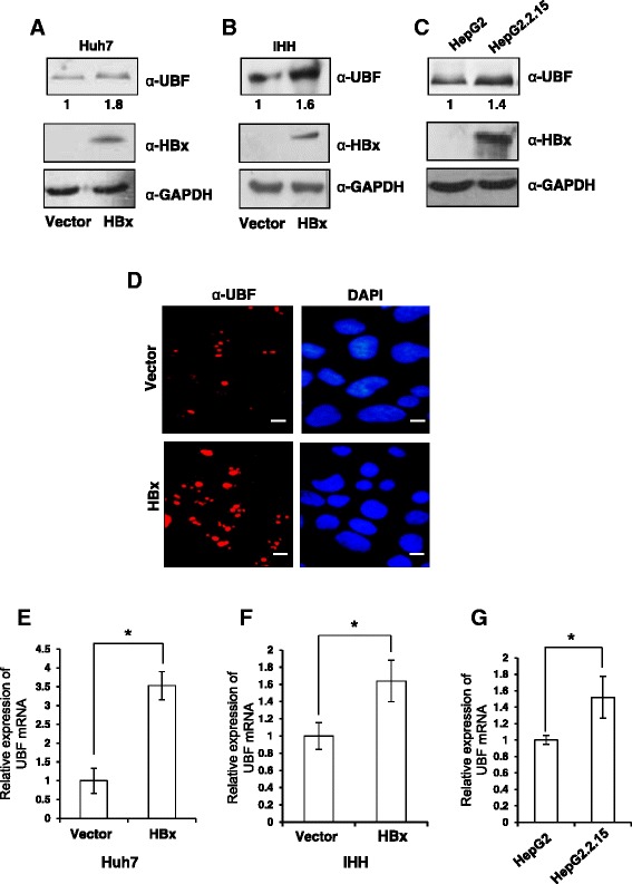
Figure 1.

Regulation of UBF expression under the HBx microenvironment. (A) & (B) Huh7 and IHH cells were transiently transfected with vector or HBx expression plasmids. After 48 h, cell lysates were western blotted for UBF, HBx and GAPDH. (C) HepG2 and HepG2.2.15 cell extracts were western blotted for UBF, HBx and GAPDH as above. (D) Huh7 cells were transiently transfected as in panel A and processed for immunofluorescence with anti-UBF antibody (Red). DAPI staining (Blue) was used to visualize nuclei. Scale bar represents 50 μm (E) & (F), Huh7 and IHH cells were transiently transfected as in panel A, B and total RNA was isolated for measuring UBF mRNA levels by RT-qPCR using primers mentioned in Additional file 1: Table S1 (G) The UBF mRNA levels in asynchronously growing HepG2 and HepG2.2.15 cells was measured as above. All data are represented as mean ± S.D of three independent experiments. * represents a statistically significant difference of p <0.05.
