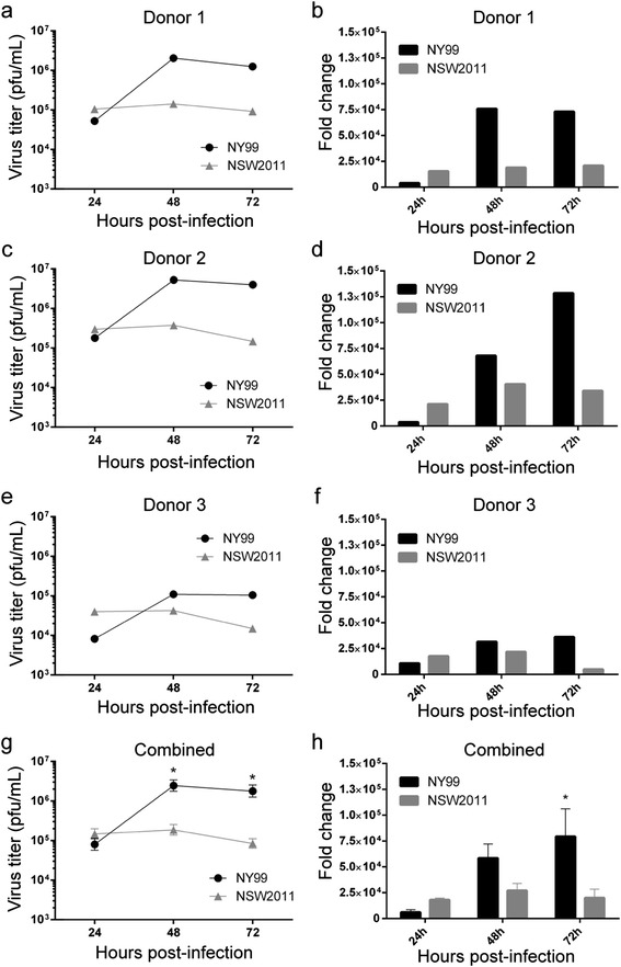
Figure 1.

Replication of WNV NY99 and WNV NSW2011 in human MoDCs. MoDCs from donors 1 (a, b), 2 (c, d) and 3 (e, f) were infected with WNVNY99 and WNVNSW2011 at MOI = 1. Cell culture supernatant was collected at 24, 48 and 72 hours post-infection and virus titer was determined by plaque assay on BHK cells (a, c, e). Total cellular RNA was collected at 24, 48 and 72 hpi and vRNA levels as determined by qRT-PCR were normalised to a combination of three endogenous controls (TBP, GAPDH and PPIA), and was expressed as fold change from uninfected samples (b, d, f). The data from all donors (donors 1 to 3) were combined and error bars represent standard error of the mean (g, h). A Student’s t-test was performed to determine statistical significance at each time point (*: P ≤ 0.05).
