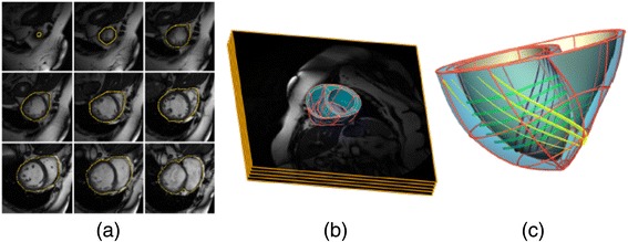
Figure 1.

Patient-specific bi-ventricular model. (a) In-vivo cardiac MRI slices showing manually segmented epicardial contour. (b) 3D cardiac model overlaid on the MRI stack. (c) Finite-element mesh with tri-cubic Hermite elements showing the main direction of fibre orientation at epicardium (yellow), midwall (green) and endocardium (purple). Reproduced with permission from [39]
