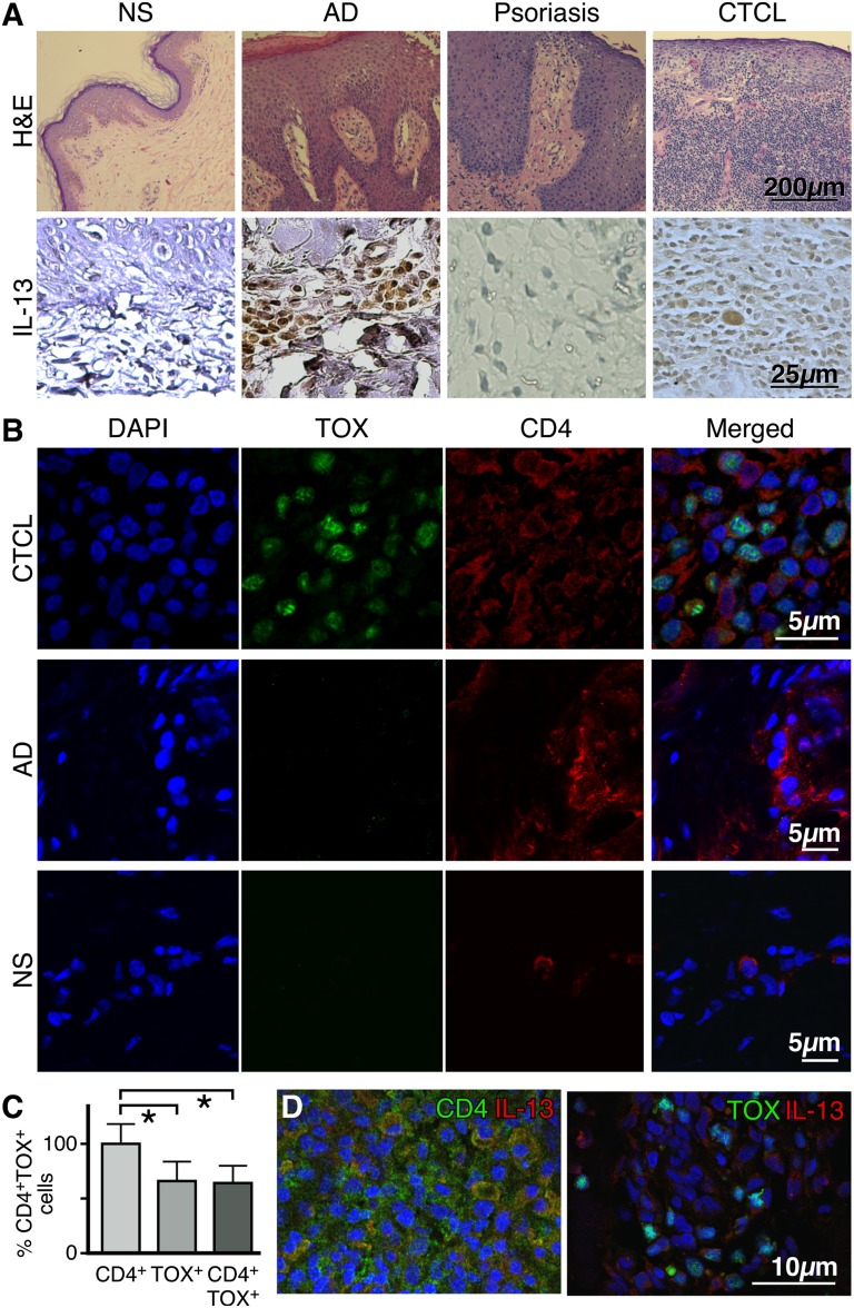
Figure 1.
IL-13 is expressed by tumor cells in the skin lesions of CTCL patients. (A, upper panel) Representative hematoxylin and eosin stains of normal skin (NS), atopic dermatitis (AD), psoriasis, and CTCL skin (original magnification ×100). (A, lower panel) Immunohistochemical staining for IL-13 expression in skin biopsies from NS (n = 3), AD (n = 5), psoriasis (n = 5), or CTCL (n = 17) (original magnification ×400). (B) Double-color immunofluorescence labeling of frozen skin samples from NS (n = 3), AD (n = 3), and CTCL (n = 7) biopsies stained with antibodies to CD4 (surface staining) and TOX (nuclear staining). A representative example is shown (original magnification ×1000). (C) Proportion of CD4+ and TOX+ cells in the skin of CTCL patients. Error bars are mean ± standard deviation (SD) (n = 7). Statistics were derived by Student t test. (D) Representative example of double-color immunofluorescence staining for CD4 and IL-13 (upper panel) or TOX and IL-13 (lower panel) (original magnification ×400). Skin samples from 7 CTCL patients were analyzed giving similar results. 4,6 diamidino-2-phenylindole stains nuclei.

