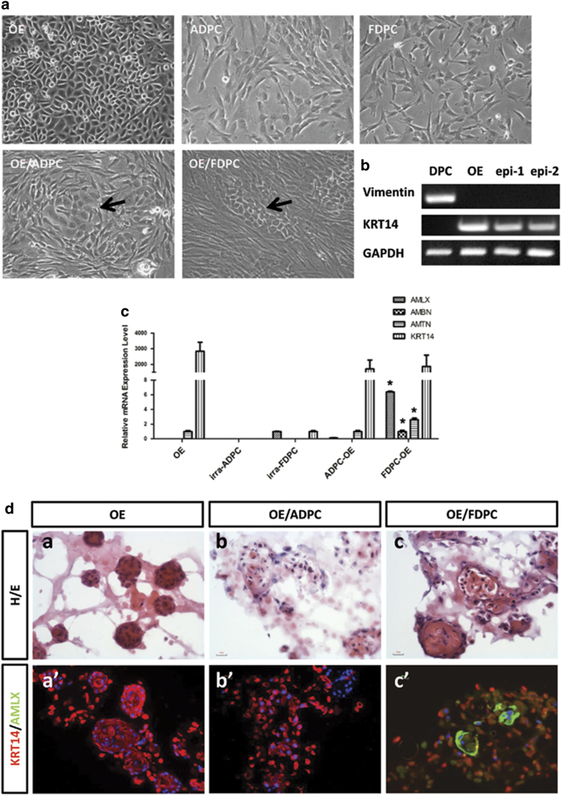
Figure 1. Oral epithelial cells (OEs) co-cultured with either fetal dental mesenchymal/papilla cells (FDPCs) or adult dental pulp cells (ADPCs).
(a): Oral epithelial cells (OEs), irradiated adult dental pulp cells (ADPCs), and irradiated fetal dental papilla cells (FDPCs) were cultured in vitro. OEs were 2-dimensionally co-cultured with either ADPCs or FDPCs (OE/ADPC and OE/FDPC). OEs co-cultured with ADPCs and FDPCs formed colonies (arrow). (b): Co-cultured OEs (epi-1 and epi-2) expressed KRT14, while no vimentin expression was observed. (c): The relative mRNA expression levels of amelogenin (AMLX), ameloblastin (AMBN), amelotin (AMTN), and cytokeratin 14 (KRT14) were detected by qPCR. OEs co-cultured with FDPCs had significantly higher expression levels of AMLX, AMBN, and AMTN than those co-cultured with ADPCs. Y-axis: fold change in gene expression. Scale bar: 50 μm. *, p < 0.01. (d): FDPCs promote the ameloblast-lineage differentiation of OEs under 3-D co-culturing conditions. OEs cultured alone under 3-D conditions in MatrigelTM formed solid cell clusters (a) that stained extensively for KRT14 but that showed no positive signal for AMLX (a’). OEs co-cultured with ADPCs under 3-D conditions in MatrigelTM failed to form epithelial nests (b). Cells were stained positive for KRT14 but not for AMLX (b’). OEs co-cultured with FDPCs under 3-D conditions in MatrigelTM formed glandular structures (c). OE cells showed positive signals for both KRT14 and AMLX (c’). Scale bar: 20 μm.

