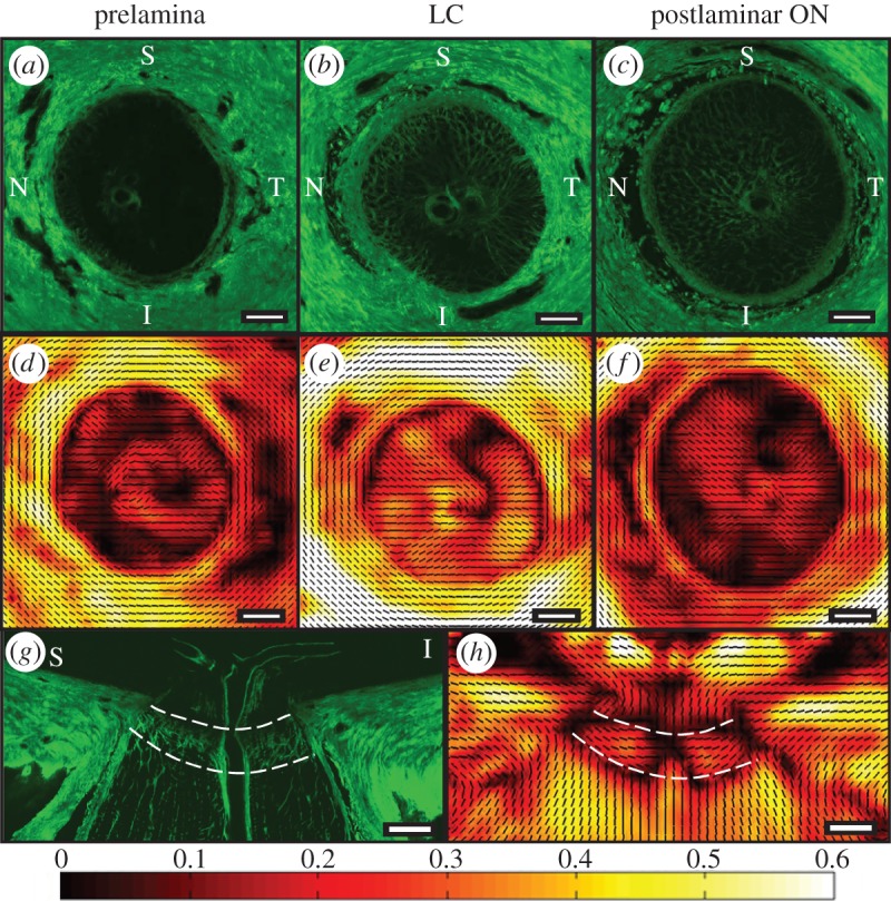
Figure 2.

(a) SHG images from transverse ONH sections acquired from the prelamina, (b) LC and (c) postlaminar optic nerve (ON) from the left eye of an 88-year-old female donor. Corresponding PFO (black PFO orientation lines at 100 µm intervals) and DOFA (colour coded from black (0: poorly aligned) to white (0.6: highest alignment)) maps of the same (d) prelamina, (e) LC and (f) postlaminar optic nerve. (g) SHG image of a longitudinal ONH section (superior–inferior orientation) from the left eye of a 78-year-old male donor and (h) its corresponding PFO and DOFA map. The anterior and posterior LC boundaries are denoted by white dashed lines. Note: PFO is not meaningful when DOFA is pure black. S, superior; N, nasal; I, inferior; T, temporal. Scale bars, 500 µm.
