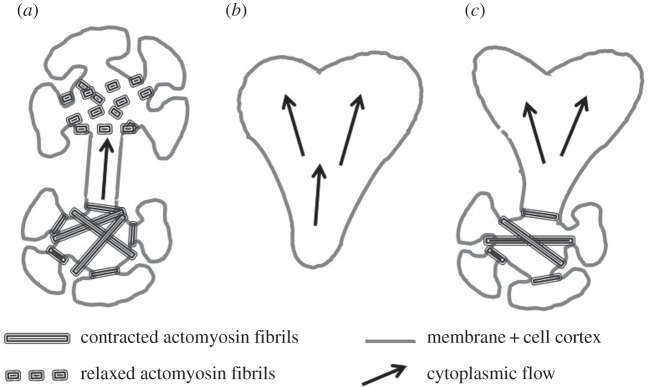
Figure 1.

Schematic drawing of the three main types of microplasmodia. The interface of each cell type consists of a cell membrane and an underlying thin actomyosin cortical layer. This layer is contractile and both its thickness and its effective tension may vary in space and time. (a) The chain type consists of several highly invaginated spherical heads connected by tube parts; these heads are highly contractile in particular due to the presence of large actomyosin fibrils which assemble (bottom head) and disassemble (top head) periodically. (b) Amoeboid type characterized by a flat shape, highly irregular and only a cell cortex. (c) Hybrid type characterized by a combination of the other two types. In each type, cytoplasmic flows (large arrows) are generated by the contractile activity of cell cortex and fibrils (adapted from Gawlitta et al. [8]).
