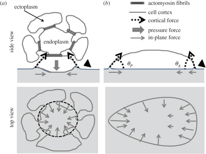
Figure 8.
Model of force transmission to the substratum for two kinds of microplasmodia. (a) Cross section of the head of a chain type microplasmodium: the invaginated ectoplasm attaches in the inner part of the contact area with a contact angle lower than 90°; cortical forces transmitted to substratum are tangential to the invagination pockets and in-plane component is inwardly directed; an internal pressure inside the pocket counterbalances the forces in the vertical direction; fibrils maintain the pocket shape and possibly enhance cortical tension. (b) Cross section of an amoeboid microplasmodium: all forces are exerted by the peripheral cortex and oriented along the cell contact profile defined by the geometrical contact angles θP and θA. The cortex force is deforming the substrate upward in both cases (arrowheads on the right of (a,b)) but this deformation was not detected.

