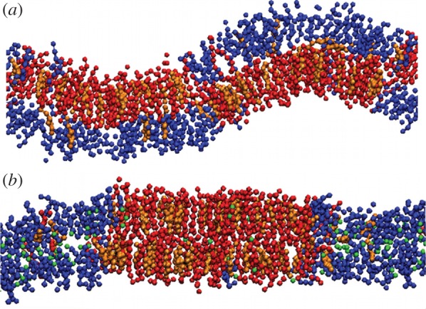
Figure 3.

(a) Snapshot in the (x, z) view for a DUPC/DSPC/chol membrane slice. Beads of different colours are used for DUPC (blue), DSPC (red) and chol (orange). Water molecules are not plotted. (b) Addition of 0.77 chlf molecules per lipid (green beads) promotes phase symmetry (registration) and as a consequence a planar membrane conformation is favoured.
