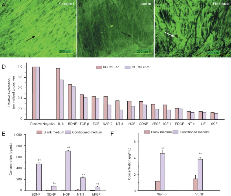
Figure 2.

hUCMSCs express and secrete extracellular matrix and neurotrophic factors that enhance nerve regeneration.
(A–C) Extracellular matrix components deposited by hUCMSCs were visualized by immunofluorescence staining. Arrows indicate positive expression. FITC was the dye. Scale bars: 200 μm. (D) Cytokine antibody array assay revealed neurotrophic factor expression. hUCMSC-1 and hUCMSC- 2 represent cultures derived from different umbilical cords. (E, F) BDNF, GDNF, HGF, NT-3, bFGF, NGF-β and VEGF protein levels were detected in hUCMSC-conditioned medium and control medium by ELISA. All data are expressed as the mean ± SD. Statistical analysis was performed using one-way analysis of variance followed by Tukey's test. **P < 0.01, vs. control (blank) medium. hUCMSCs: Human umbilical cord-derived mesenchymal stem cells; IL-6: interleukin-6; BDNF: brain-derived neurotrophic factor; TGF-β: tumor growth factor-β; EGF: epidermal growth factor; NAP-2: neutrophil activating protein-2; NT-3: neurotrophin-3; HGF: hepatocyte growth factor; GDNF: glial-derived neurotrophic factor; VEGF: vascular endothelial growth factor; IGF-1: insulin-like growth factor-1; PDGF: platelet-derived growth factor; NT-4: neurotrophin-4; LIF: leukemia inhibitory factor; SCF: stem cell factor; bFGF: basic fibroblast growth factor; NGF-β: nerve growth factor-β; ELISA: enzyme linked immunosorbent assay.
