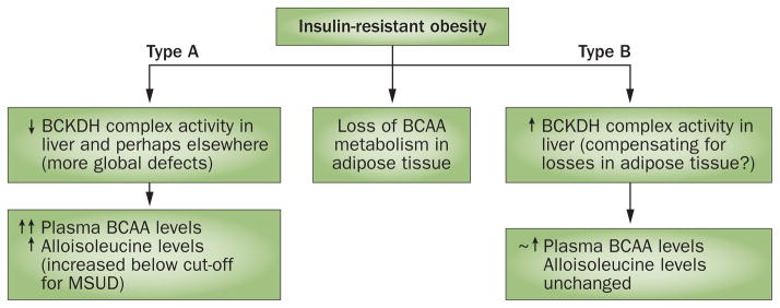
Figure 5.

Patterns of altered BCAA metabolism observed in animal models of obesity. Losses of adipose tissue BCAA metabolic gene and protein expression in obesity have consistently been observed. In a rat model of diet-induced obesity,63 reduced levels of BCAA metabolizing enzymes in adipose tissue seem to be compensated for by increased hepatic BCKDH activity163 (termed a type B response). In contradistinction, hepatic BCKDH was also attenuated in other models such as ZDF rats,161,162 Zucker fa/fa rats72,73, ob/ob mice72 and Otsuka Long–Evans Tokushima Fatty rats162 (termed a type A response). Multiple peripheral tissues were examined and found to be affected in Zucker rats.73 These distinct phenotypes are important because uncompensated loss of BCAA metabolism in multiple peripheral tissues could result in a higher range of plasma BCAAs that, when observed in states of obesity, associate to a greater extent with insulin resistance, levels of glycaemia and future T2DM. Alloisoleucine elevations below the level used to screen for MSUD have been proposed as a strategy to distinguish between these phenotypes.75 Levels of BCAAs were considerably increased in models in which metabolism was impaired (↑↑). Abbreviations: BCAA, branched-chain amino acid; BCKDH, branched-chain α-keto acid dehydrogenase; MSUD, maple syrup urine disease. Adapted with permission from John Wiley and Sons © Olson, K. C. et al. Obesity (Silver Spring) 22, 1212–1215 (2014).75
