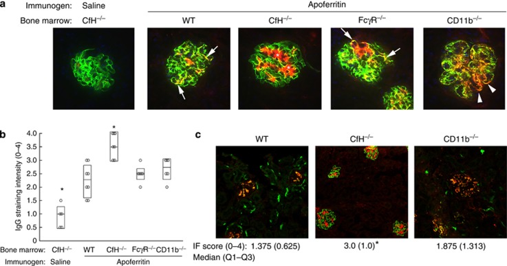
Figure 2.
Glomerular immune complex deposition in chronic serum sickness (CSS). (a) Representative immunofluorescence photomicrographs from complement factor H (CfH)−/− mice chimeric for wild-type (WT), CfH−/−, FcγR−/−, and CD11b−/− bone marrow, or (c) native WT, CfH−/−, and CD11b−/− mice (i.e., without bone marrow transfer) actively immunized with apoferritin to induce chronic serum sickness, or saline as control. Dual staining for C3 (green) and IgG (red) is shown; the yellow color indicates immune complexes containing IgG and C3. Because animals lacked plasma CfH, there was strongly positive glomerular C3 staining in all groups. The asterisks and arrows depict mesangial and peripheral capillary wall immune complex deposits, respectively. The arrowheads show the more granular peripheral capillary wall immune complex staining in CD11b−/− chimeras. (b, c) Semiquantitative scoring for IgG staining intensity from individual mice from all groups. In b, data are shown as median (horizontal line), and Q3-Q1 in the boxes, whereas in c these are presented below the figures. Group medians were significantly different (P=0.02, Mood Median test). *P<0.05 versus all other groups.

