
Figure 1.
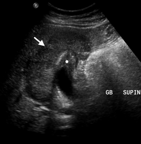
Ultrasound of GB: Well defined, irregular heterogeneous mixed hypo- and iso-echoic mass at the gallbladder fossa (arrow) burst the overlying liver parenchyma and with direct invasion of gallbladder wall at fundus (white dot), giving the possibilities of either of hepatic or gallbladder origin.