Figure 3.
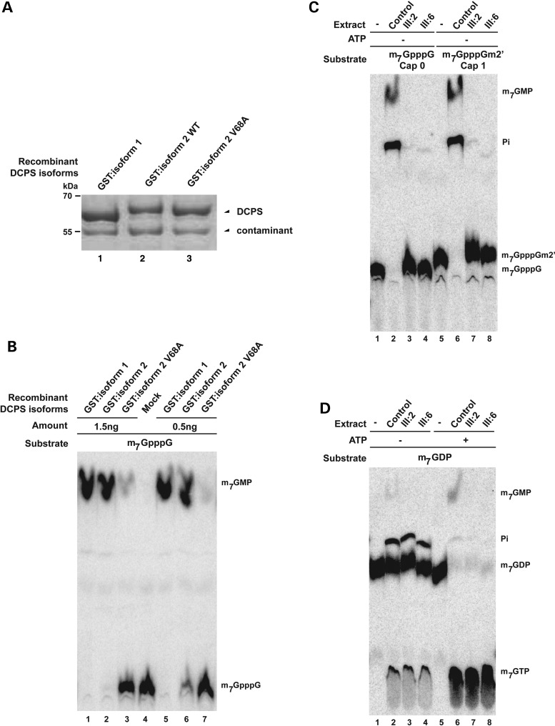
Patients' cells have no detectable enzymatic DCPS activity. (A) Protein profiles of purified recombinant GST:DCPS isoform 1 and GST:DCPS isoform 2 and isoform 2 V68A on SDS–PAGE. The contaminant migrating below GST-DCPS serves as a loading control. Positions of migration of molecular weight markers are indicated on the left side. (B) Enzymatic assay demonstrates the reduced activity of the DCPS when 7 amino acids is inserted between exon 1 and exon 2 of DCPS protein, especially in the V68A mutant. (C) Enzymatic assay demonstrates the loss of DCPS activity in patient's cells using m7GpppG and m7GpppGm2′ substrates. Purified substrates are presented in lanes 1 and 5. (D) In vitro assays demonstrate that the absence of DCPS-dependent conversion of m7GTP in extracts of cells from two affected individuals. m7GTP is efficiently formed from m7GDP in the presence of ATP (lanes 5–8), demonstrating that all extracts are similarly active. Trace of ATP in extract explains the residual formation of m7GTP in the absence of added ATP (lanes 1–4). The purified substrate (+/− ATP) is presented in lanes 1 and 5.

