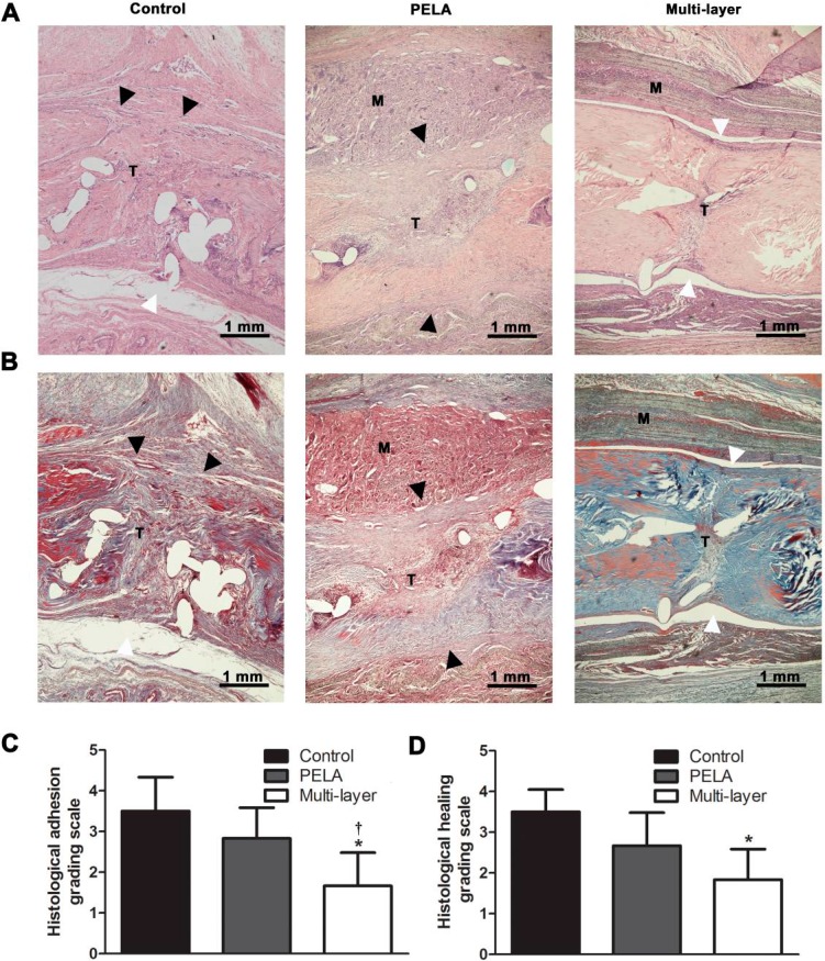
Figure 3.
HE (A) and Masson (B) staining of untreated repair site, repair sites wrapped with unloaded PELA fibrous membrane and wrapped with a multi-layer fibrous membrane. White arrowheads indicate the interface without peritendinous adhesions, while black arrowheads indicate peritendinous adhesions between the membrane (M) and tendon (T) (Scale bar = 1 mm). Peritendinous adhesions and tendon repair are evaluated by determining histological adhesion grade (C) and histological healing grade (D). * p < 0.05 compared with control group; † p < 0.05 compared with PELA membrane group. Data are expressed as mean ± SEM for six tendons/groups.

