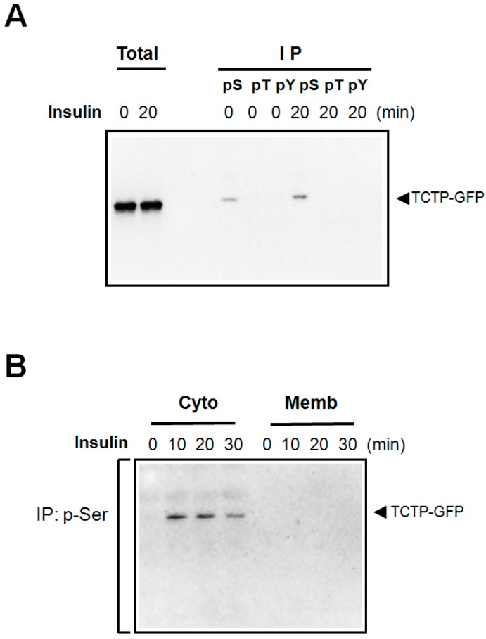
Figure 1.
Insulin-induced phosphorylation of TCTP in 293T cells and the cytosolic localization of phosphorylated TCTP. (A) Following starvation, pEGFP-N1-TCTP-transfected 293T cells were treated with insulin at 100 nM concentrations for indicated times. Cells were harvested, and subsequently lysed in the lysis buffer containing detergent (1% Triton X-100) on ice. After brief sonication, samples were centrifuged at a 10,000 rpm for 10 min. Resulting supernatants were used as total cell lysates. For immunoprecipitation, anti-p-Ser-, -p-Tyr-, and -p-Thr-specific antibodies were used to precipitate the phosphorylated TCTP in cell lysates. The immune complexes so obtained were resolved in SDS-PAGE and then immunoblotted using anti-GFP-antibody; (B) Following transfection of pEGFP-N1-TCTP construct, 293T cells were treated with 100 nM insulin. Cell lysates thus obtained were fractionated into membrane and cytosolic fractions, as indicated in the Materials and Methods sections. Each fraction was then immunoprecipitated using anti-p-Ser antibodies, followed by immunoblotting with anti-GFP antibody.

