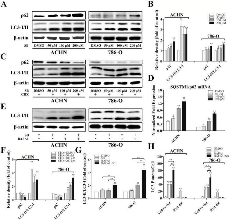
Figure 2.
Silibinin induces autophagic flux in RCC ACHN and 786-O cells. (A) Cells were treated with indicated dose of silibinin (SB) for 24 h, the levels of p62 and LC3-I/II were checked by Western blot and quantitative analyses were done (B); (C,F) RCC cells were treated with the indicated dose of silibinin for 24 h in the presence and absence of 350 nM cyclohexamide and the levels of p62 and LC3-I/II were examined; (D) The expression of SQSTM1/p62 mRNA accessed by real-time PCR. RCC cells were treated with 50 μM of silibinin for 24 h in the presence and absence of 10 nM BAFA1 and the levels of LC3-I/II (E,G) and the numbers of LC3 puncta (H) were examined. Blots are representative of three separate experiments. Error bars represent SDs. * p < 0.05; ** p < 0.01.

