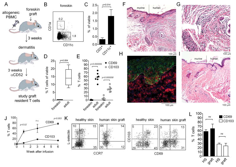
Figure 1. Skin T cells in a human engrafted mouse model recapitulate T cell populations in adult human skin.
(A) A human engrafted mouse model was used to discriminate resident from recirculating T cells in skin. NSG mice were grafted with human foreskin and injected i.v. with allogeneic human PBMC, and alemtuzumab (αCD52) was used to deplete recirculating human T cells from grafted human skin. (B–E) human foreskins contained resident APC but few T cells. (B) Flow cytometry staining of collagenase digested foreskin demonstrated both CD11c+ DC and CD1a+ LC were present in foreskin. (C) Approximately four-fold more DC than T cells were present in foreskin, as assayed by the % viable cells of each cell type in collagenase digested foreskin. (D) T cells were 45-fold more frequent in healthy adult human skin than in foreskins; the percentage of CD3+ T cells in collagenase digested foreskin versus adult skin is shown. (E) The few T cells present in human foreskin lacked markers characteristic of TRM. The percent of T cells expressing each marker in collagenase treated foreskin and adult skin are shown. The mean and SEM of four foreskins (C–E) and seven adult skin donors (D,E) are shown (test: T-test). (F–H) allogeneic PBMC injected i.v. into grafted mice migrated specifically into the human skin graft and induced an inflammatory dermatitis. Images are representative of eight PBMC injected and eight saline injected mice. (F) H&E stained section demonstrating the junction between mouse and human skin in grafted mice. (G) H&E stain, 20X view of inflammatory dermatitis. (H) A cryosections of human grafted skin immunostained for CD3 (red) and keratin (green) are shown. T cells were evident in both the dermis and the epidermis. The dermoepidermal junction is indicated by a white line. (I) in the absence of infused allogeneic PBMC, no inflammatory infiltrate was observed and few or no T cells were visualized in skin. (J) CD69 up-regulation preceded CD103 up-regulation in T cells migrating into human skin grafts. Human skin grafts were harvested at the indicated time points, collagenase digested, and T cells were analyzed for CD69 and CD103 expression by flow cytometry. The mean and SEM of two grafted mice per time point are shown (test: T-test). (K,L) T cell populations in grafted skin recapitulate those observed in adult human skin. T cell expression of TCM (CCR7/L-selectin) and TRM (CD69/CD103) markers as assayed by flow cytometry are shown. T cells were isolated by collagenase digestion from healthy adult human skin or from skin grafts on PBMC infused mice. Representative histograms (K) and mean and SEM of expression of CD69 and CD103 are shown. The mean and SEM of three grafts and seven healthy skin donors are shown (test: T-test).

