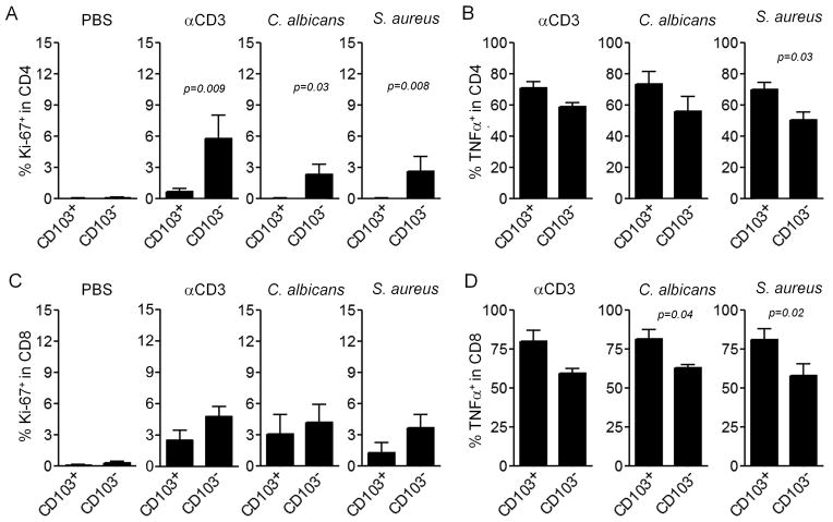
Figure 5. CD103+ TRM have a lower proliferative capacity but increased effector function.
Skin explants were injected with PBS, stimulatory anti-CD3and anti-CD28 antibodies (αCD3), or heat killed extracts of C. albicans and S. aureus. Two weeks later, T cells that had migrated out of the skin were collected and the proliferation of CD4+ TRM (A) and CD8+ TRM (C) were assayed by staining for Ki-67 and flow cytometry analysis. TNFα production was assayed by intracellular cytokine staining and flow cytometry analysis after stimulation with PMA and ionomycin. Results for CD4+ TRM (B) and CD8+ TRM (D) are shown. Results are shown for CD69+/CD103+ (CD103+ TRM), and CD69+/CD103− (CD103− TRM) The mean and SEM of three donors (PBS) and six donors (all other conditions) are shown (test:T-test).

