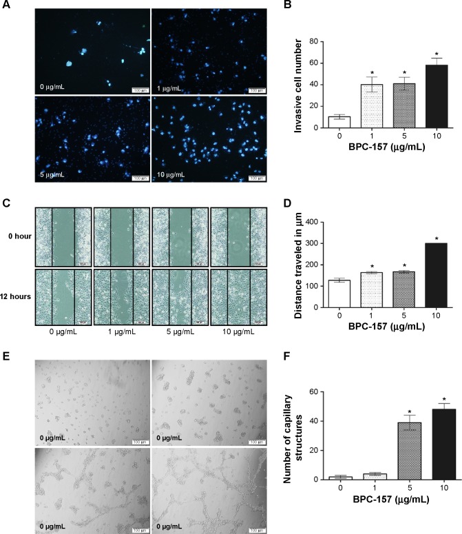
Figure 5.
Effect of BPC-157 on cell migration and tube formation of HUVECs.
Notes: (A) Representative photomicrographs showing the stained cells on the lower side of membranes. The cells in the upper chambers were treated with DMSO (control, 0 μg/mL) or 1 μg/mL, 5 μg/mL, and 10 μg/mL BPC-157. After 12 hours of incubation, those cells which had migrated to the lower chambers were stained and the numbers were counted. (B) Quantification of cell migration in HUVECs is shown. (C) A scratch wound was created using a 20 μL sterile serological pipette in a confluent HUVEC culture after incubation with BPC-157. The images were taken at 0 hours and 12 hours. The black lines show the area where the scratch wound was created. (D) Quantification of cell motility in HUVECs is shown. (E) Effect of BPC-157 on tube formation in HUVECs. Photomicrographs represent the matrigel tube formation of HUVECs after 8 hours’ incubation. (F) Quantification of capillary structure in HUVECs is shown. Data are presented as mean ± SD. Differences between the treated and untreated control groups were determined by Student’s unpaired t-test. *P<0.05 significantly different from the control group.
Abbreviations: BPC-157, body protective compound 157; DMSO, dimethyl sulfoxide; HUVECs, human umbilical vein endothelial cells; SD, standard deviation.

