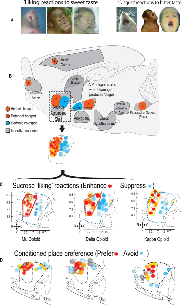
Figure 1. Causal hedonic hotspots and coldspots in the brain.
A) Top shows positive hedonic orofacial expressions (‘liking’) elicited by sucrose taste in rat, orangutan, and newborn human infant. Negative aversive (‘disgust’) reactions are elicited by bitter taste. B) shows sagittal view of hedonic hotspots in rat brain containing nucleus accumbens, ventral pallidum, and prefrontal cortex. Hotspots (red) depict sites where opioid stimulation enhances ‘liking’ reactions elicited by sucrose taste. Coldspots (blue) show sites where the same opioid stimulation oppositely suppresses ‘liking’ reactions to sucrose. C) Nucleus accumbens blow-up of medial shell shows effects of opioid microinjections in NAc hotspot and coldspot. (red/orange dots in hotspot = >200% increases in ‘liking’ reactions; blue dots in coldspot = 50% reductions in ‘liking’ reactions to sucrose). Panels show separate hedonic effects of mu opioid, delta opioid and kappa opioid stimulation via microinjections in NAc shell on sweetness ‘liking’ reactions. Bottom row shows effects of mu, delta or kappa agonist microinjections on establishment of a learned place preference (i.e., red/orange dots in hotspot) or place avoidance (blue dots). Surprisingly similar patterns of anterior hedonic hotspots and posterior suppressive coldspots are seen for all three major types of opioid receptor stimulation. Modified from (Castro and Berridge, 2014).

