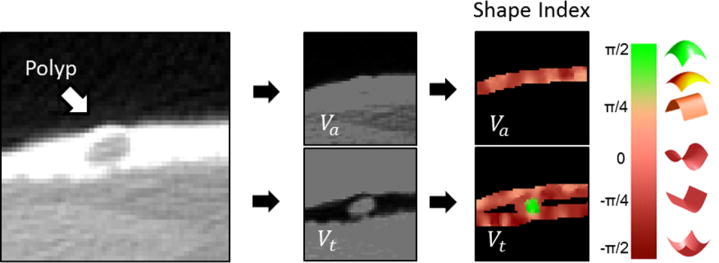
Figure 3.

In context-specific detection, the CTC image volume is divided into a separate air-filled lumen image volume (Va) and a lumen image volume that represents regions covered by fecal tagging (Vt). The initial detection of suspicious sites is performed independently on both volumes. High values of the SI feature of Vt (green color) indicate the location of a polyp.
