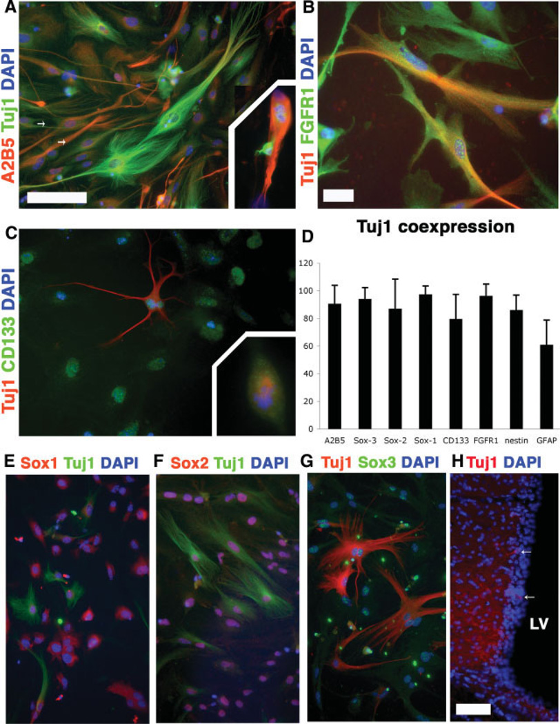
Figure 4.
Class III β-tubulin/Tuj1 labels putative neural stem cell (NSC)/progenitor populations. Multiple antibody labeling in undifferentiated passage 3 subventricular NSCs revealed coexpression of A2B5 ([A], arrows), FGFR1 (B), and CD133/Prominin-1 (C). Tuj1 was expressed in the mitotic spindle of dividing cells ([A, C], insets). (D): Substantial fractions of Tuj1+ cells expressed NSC and progenitor markers. CD15 and NG2 were examined and were not expressed in undifferentiated cultures. (E–G): Tuj1 was expressed in multiple members of the Sox protein family, as well as in the supependymal cell layer of the lateral ventrical (H). Coronal section of the subventricular zone revealed that Tuj1+ cells were present in the subventricular layer adjacent to the LV (arrows). Scale bars = 50 µm (A, C, E–G), 25 µm (B), and 100 µm (H). Abbreviations: DAPI, 4,6-diamidino-2-phenylindole; FGFR1, fibroblast growth factor receptor 1; GFAP, glial fibrillary acidic protein; LV, lateral ventricle.

