
Figure 6.
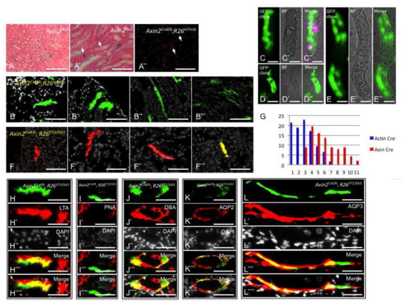
Wnt-Responsive Cells (WRCs) are clone forming cells and lineage-restricted in-vivo. Sections from AxinlacZ/lacZ kidneys stained with beta-galactosidase and counter-stained with eosin showing Axin2 expression within the collecting system and proximal tubules (A, A′). Axin2CreER;R26mTmG kidneys that were pulsed for 4 days show GFP expression within the collecting system and proximal tubules (A″, white arrows). (B-B‴) Axin2CreER;R26mTmG kidneys that were pulsed from E17.5 up to the 3rd postnatal month show large GFP+ clones within proximal tubules and collecting ducts. (C–D) Intact PT segments from Axin2CreER;R26mTmG kidneys show large GFP+ clones confined to individual segments. Axin2CreER; R26VT2/GK3 kidneys that were pulsed from E17.5 up to the 5th postnatal month show singly colored large clones within proximal tubules (F, F′) and collecting ducts (F″, F‴). (G) Histogram which graphically represent the difference in clonal outcomes of all cells (blue) vs. WRCs (red). (E,F) Clones derived from WRCs are lineage restricted to either PT or CD fates. Within the cortex, clones are LTA+, PNA− indicating PT fate. (G–I) Within the renal papilla, clones are DBA+, AQP2+, AQP3+ indicating CD fate. PT, proximal tubule; DT, distal tubule, CD, collecting duct. Scale bar: A′, A″ (50μm); A, B-B‴, F-F‴, C–E (25μm).