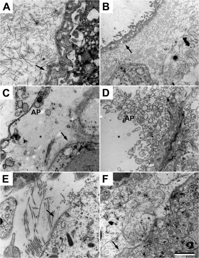
Fig 5. A1M prevents the placental damages caused by free HbF.

Transmission electron microscopy analysis of placental tissue. (A-B) Placental tissue from control animals. Normal extracellular matrix with dense bundles of collagen fibers and a normal electron dense barrier (arrow) can be seen. (C-D) Cell-free HbF causes severe damage to the extracellular matrix with significant loss of collagen fibers, increased numbers of extracellular apoptotic bodies (AP), cell debris, disruption of the electron dense barrier and numerous areas of empty extracellular space (star). (E-F) A1M treatment significantly normalized the structural damages. Note the normal bundles of collagen fibers, normal electron dense barrier and reduced numbers of apoptotic bodies in the extracellular space. Scale bar 1 μm.
