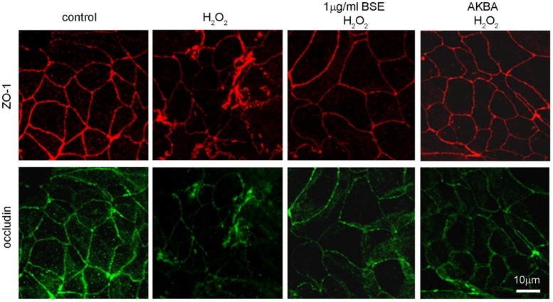
Fig 5. BSE and AKBA ameliorate alterations following exposure to H2O2 occludin and zonula occludens (ZO1) TJ proteins in Caco-2 cell monolayers.
Double immunostaining showing the distribution of ZO-1 (red) and occludin (green). Images were collected by confocal laser-scanning microscope and are representative of 3 independent experiments. Scale bar = 10 μm.

