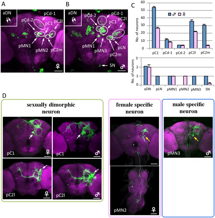
Fig 3. Sex differences in dsx-GAL4-expressing neurons.
(A, B) Posterior view of a female (A) and male (B) brain in the flies expressing UAS-mCD8::GFP under the control of dsx GAL4 (G). Islets in A and B are shown in anterior view. The genotype of flies used is y hs-flp;G13 UAS-mCD8::GFP;dsx GAL4 (G). (C) The number of neurons contained in 11 dsx-GAL4-expressing neuron clusters was compared between the female and male brain. Values represent the mean ± s.e. (n = 12). (D) Examples of sex differences in dsx-GAL4-expressing neuron clusters. The somata of neuron clusters and single neurons indicated as MARCM clones are shown using white and yellow arrows, respectively. The brains were stained with anti-GFP (or anti-mCD8) antibodies (green) and nc82Mab (magenta). The scale bars represent 50 μm.

