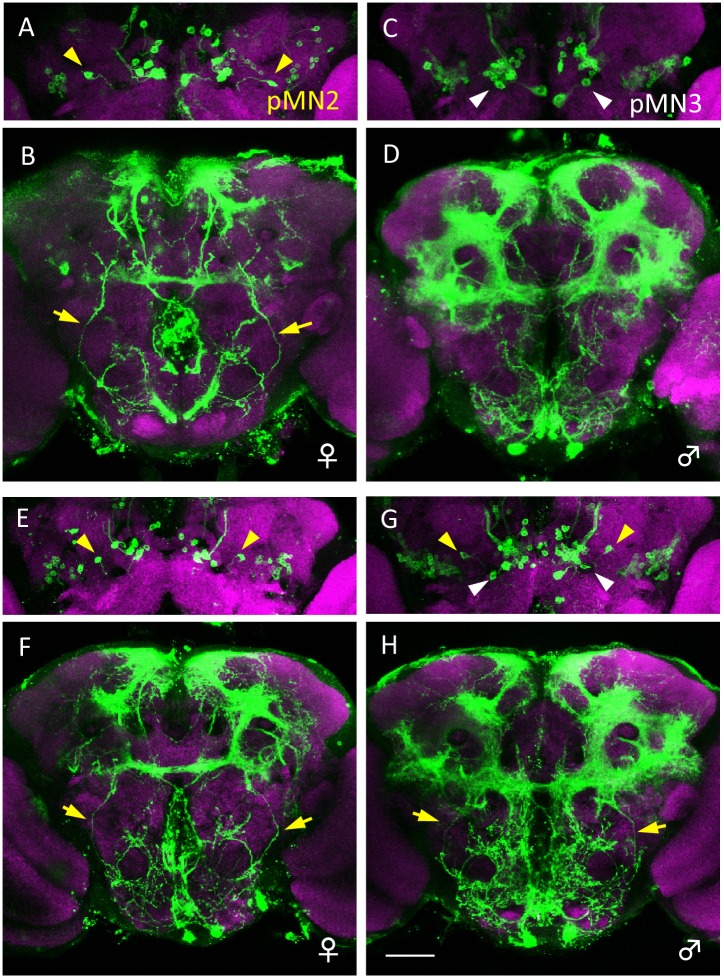
Fig 5. Ectopic formation of female-specific pMN2 in the male brain by artificial expression of the cell death inhibitor p35 in dsx-expressing neurons.
(A, B) A pair of cell bodies of female-specific pMN2 neurons (yellow arrowheads in A) and their neurites (yellow arrows in B) are present in a wild-type female of y hs-flp/+; UAS-mCD8::GFP/+; dsx GAL4 (G)/+. (C, D) A pair of cell bodies of male-specific pMN3 neurons (white arrowheads in C) are present in a wild-type male of y hs-flp/Y; UAS-mCD8::GFP/+; dsx GAL4 (G)/+. (E-H) A pair of cell bodies of pMN2 (yellow arrowheads in G) and its neurites (yellow arrows in H) are labeled together with pMN3 neurons (white arrowheads in G) in the brain of a male fly in which cell-death has been blocked. The fly genotype is y hs-flp/Y; UAS-mCD8::GFP/UAS-p35; dsx GAL4 (G)/+. In the female of y hs-flp/+; UAS-mCD8::GFP/UAS-p35; dsx GAL4 (G)/+, the cell bodies of pMN2 neurons and their neurites are observed (yellow arrowheads in E and yellow arrows in F, respectively). Brains were doubly stained with anti-GFP (green) and nc82 mAb (magenta). The scale bar represents 50 μm.

