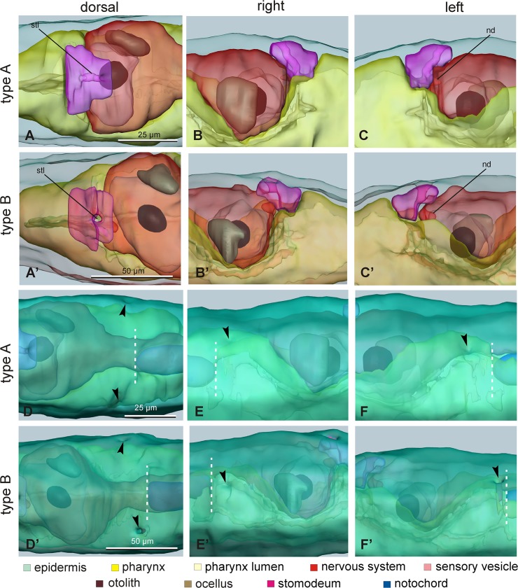
Fig 8. Details of 3D reconstructions of larvae.
A-C’. Detail of the stomodeum (shocking pink) and its relationships with the pharynx (yellow) and the neurohypophyseal duct in larvae of type A (A-C) and type B (A’-C’), viewed from dorsal (A, A’), right (B, B’) and left (C, C’) sides. nd: neurohypophyseal duct; stl: stomodeum lumen; green: epidermis, pale yellow: pharynx lumen, red: nervous system, pink: sensory vesicle lumen; brown: otolith; pale brown: ocellus. D-F’. Detail of posterior region of cephalenteron to compare the position of the two atria (arrowheads) and the anterior end of the notochord (white dotted line) in larvae of type A (D-F) and type B (D’-F’), viewed from dorsal (D, D’), right (E, E’) and left (F, F’) sides. The epidermis (green) has been intensely colored to allow visualization of atria; internal structures are semi-hidden. Enlargements are the same in each row of pictures (see the scale bars in the first column).

