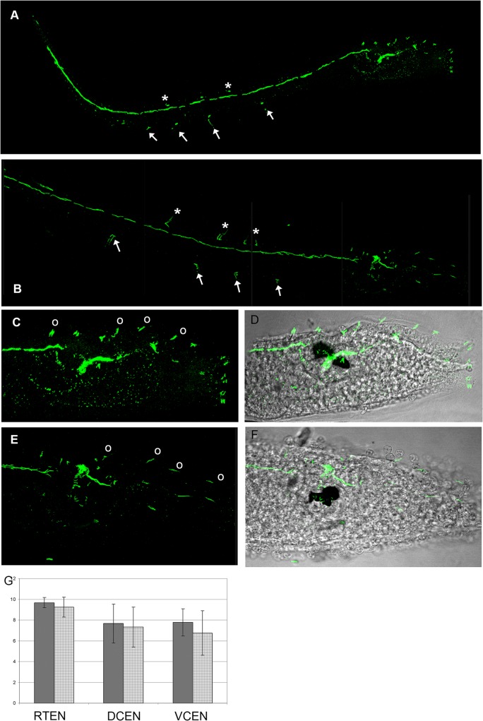
Fig 9. Immunolabeling of nervous fibres in late larvae of type A (A,C,D) and type B (B,E,F).
A,B confocal laser microscope imagines of whole larvae. Asterisk indicate dorsal caudal epidermal neurons, arrows indicate ventral caudal epidermal neurons. C,E. Magnifications of the trunk regions. Circles indicate trunk epidermal neurons. D,F. Superimposition of C and D with transmission microscope images. G. Graph showing the mean number of neurons ± standard deviation. DCEN: dorsal caudal epidermal neurons of the tail; VCEN ventral caudal epidermal neurons of the tail; TEN: trunk epidermal neurons.

