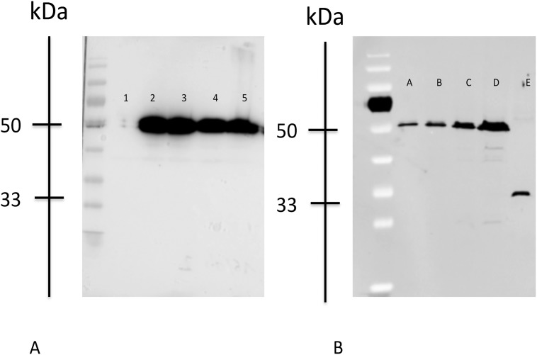
Fig 2. Western Blot.
Western blot analysis of wild-type and mutant LHX4 proteins from transfected heterologous human embryonic kidney 293T cells. The migration positions of protein standards (kDa) are shown. Control is transfection with empty vector. Lanes are identified as follows: Fig. A; 1, pcDNA3.1 (empty vector); 2, LHX4 WT-myc; 3, DelK242-myc; 4, N271S_myc; 5, Q346R-myc; Fig. B; A: LHX4 WT-myc diluted to 1/8; B: LHX4 WT-myc diluted to ¼; C: LHX4 WT-myc diluted to ½; D: LHX4 WT-myc; E: LHX4 203-myc.

