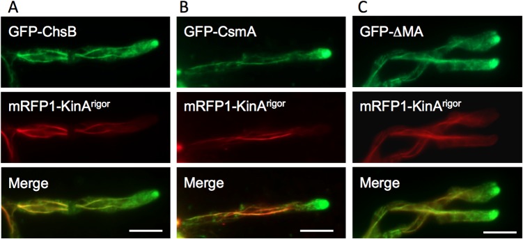
Fig 6. Colocalization of GFP-ChsB, GFP-CsmA, and GFP-ΔMA with mRFP1-KinArigor along microtubules.

(A, B) GFP-ChsB (A) and GFP-CsmA (B) localized to large accumulations at the subapical tips similar to those in the ΔkinA strains, and were observed along microtubules decorated with mRFP-KinArigor throughout the hyphae. (C) GFP-ΔMA (CsmA without the MMD) also localized to large accumulations at the subapical tips and along microtubules decorated with mRFP-KinArigor. (A-C) These strains were grown in MMGlyuu overnight. Scale bars represent 5 μm.
