
Fig. 5.
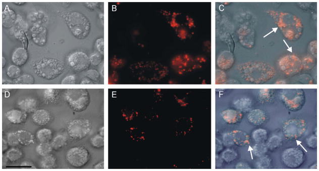
Light microscopy of live BME26 cells after uptake of hemoglobin conjugated to rhodamine. (A, D) DIC microscopy; (B, E) fluorescence microscopy; (C, F) merged images. Incubation conditions were: (A–C) cells exposed to 20 μmol l−1 rhodamine–hemoglobin, presenting labeling in small vesicles (arrows in merged image); (D–F) cells exposed to 20 μmol l−1 rhodamine–hemoglobin plus 2 mmol l−1 unlabeled hemoglobin, labeling is maintained in small vesicles (arrows in merged image). Scale bar = 20 μm.