
Figure 3. Loss of p47phox improves adriamycin-induced matrix deposition.
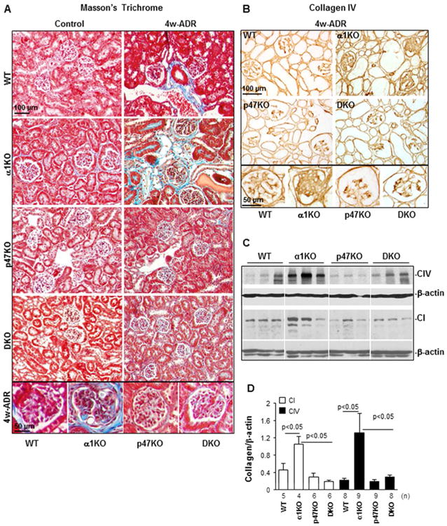
(A) Masson's Trichrome staining of kidneys from uninjured (Control) or adriamycin-treated (4w-ADR) WT, α1KO, p47phoxKO, and DKO mice. Note the presence of fibrillar collagen (blue) in both glomeruli and tubules of integrin α1KO mice which was reduced in kidneys of p47phoxKO and DKO mice. (B) Collagen IV staining of kidney sections from 4 weeks injured WT, α1KO, p47phoxKO and DKO mice. Note the high levels of glomerular collagen IV in integrin α1KO mice which were reduced in kidneys of p47phoxKO and DKO mice. (C) Equal amount of kidney lysates (20 μg/lane) from WT, α1KO, p47phoxKO, and DKO mice (n=3 shown) 4 weeks after adriamycin injection were analyzed by Western blot for levels of collagen IV and collagen I. (D) The collagen I, collagen IV and β-actin bands were analyzed by densitometry analysis and the levels of collagen IV are expressed as collagen I/β-actin or collagen IV/β-actin ratio. The values represent the mean ± SD of the number of kidneys indicated.