
Figure 4. Loss of p47phox improves adriamycin-induced oxidative stress.
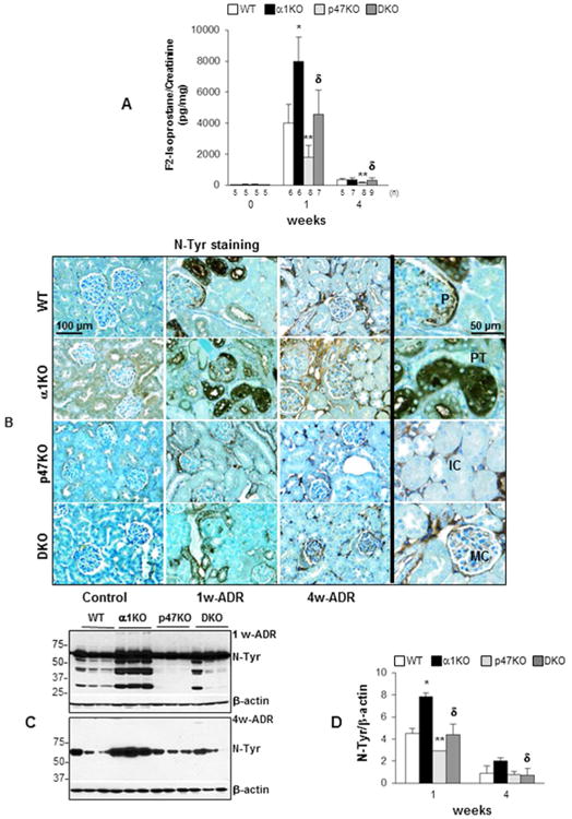
(A) The levels of urinary F2-isoprostane in the mice indicated were analyzed by ELISA before or after adriamycin injection and were expressed F2-isoprostane/urine creatinine ratio. Values are the mean ± SD of the number of mice indicated. (*), (**) and (δ) are as in Fig. 1. (B) Paraffin sections of kidneys from uninjured (Control) or adriamycin-treated (1w-ADR and 4w-ADR) WT, α1KO, p47phoxKO, or DKO mice were stained with anti-nitrotyrosine (N-Tyr) antibodies to evaluate the degree of oxidative stress-induced tyrosine nitration (brown staining). Slides were counterstained with toluidine blue (blue staining). The high-magnification images on the right show nitrotyrosine staining in infiltrating cells (IC), mesangial cells (MC), podocytes (P), and proximal tubules (PT). (C) Equal amount of kidney lysates (20 μg/lane) from WT, α1KO, p47phoxKO, and DKO mice (n=3) 1 and 4 weeks after adriamycin injection were analyzed by Western blot for levels of nitro-tyrosine. (D) The nitro-tyrosine and β-actin bands were analyzed by densitometry analysis and the levels of tyrosine nitration are expressed as N-Tyr/β-actin ratio. The values represent the mean ± SD of 3 kidneys/genotype. (*), (**) and (δ) are as in Fig. 1.