
Figure 5. Analysis of p47phox levels and localization in kidneys from uninjured and adriamycin-injured mice.
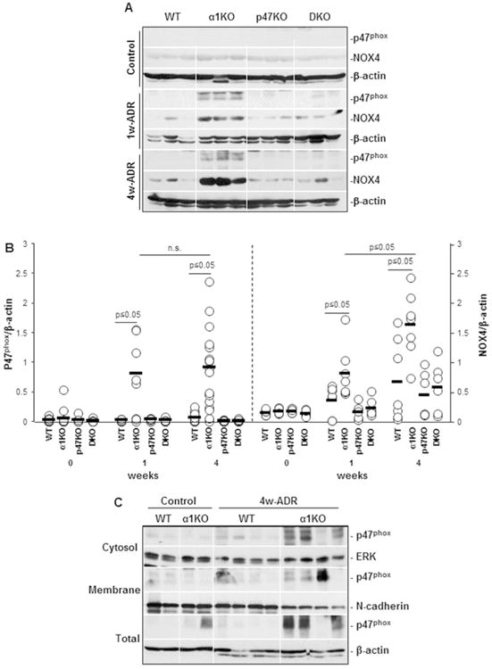
(A) Equal amount of kidney lysates (20 μg/lane) from uninjured (Control) or adriamycin-treated (1w-ADR and 4w-ADR) WT, α1KO, p47phoxKO, and DKO mice (n=3 shown) were analyzed by Western blot for levels of p47phox and NOX4. (B) The p47phox, NOX4 and β-actin bands were analyzed by densitometry and the levels of p47phox and NOX4 are expressed as p47phox/β-actin or NOX4/β-actin ratio. Open circles represent values of individual kidneys, while the bar represents the mean value. (C) Equal amount of kidney lysates (Total) membrane enriched (Membrane) and membrane deprived (Cytosol) fractions (40 μg/lane) from uninjured (Control) or adriamycin-treated (4w-ADR) WT and integrin α1KO mice (n=3 shown) were analyzed by Western blot for p47phox localization. Membranes were re-blotted with anti-ERK, anti-N-cadherin, and anti β-actin antibodies to verify the purity of various fractions.