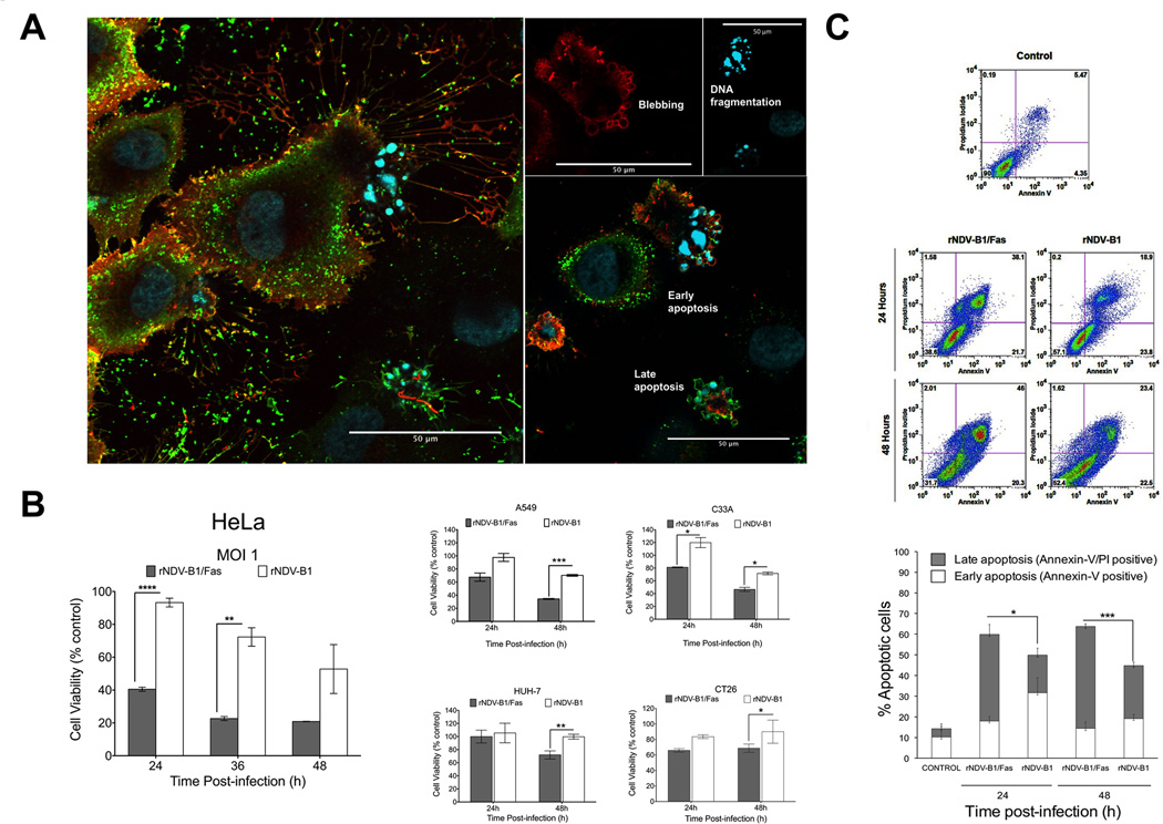
Figure 2. rNDV-B1/Fas infection induces higher cytotoxicity and an earlier apoptotic response in cancer cells.
A, cytopathic effect due to rNDV-B1/Fas infection. Confocal microscopy images of HeLa cells infected with rNDV-B1/Fas. Cells were infected at an MOI of 1 PFU/cell, fixed 20 hours post-infection and stained with monoclonal anti-human Fas antibody (red), polyclonal anti-NDV serum (green) and Hoechst for nuclear contrast. Left panel: composite Z-stack of six optical slices showing membrane and intracellular distribution of Fas receptor. Left panels show different late apoptotic feautures, like membrane blebbing and DNA fragmentation, observed among the infected population. Scale bar 50µm. B, cytotoxicity. HeLa, A549, HUH-7, C-33A and Ct26 cells were infected at an MOI of 1 PFU/cell and their viability was determined by MTT viability assay at different time points (24, 36 and 48 hours post-infection) (n=3, *p<0.05 **p<0.005, ***p< 0.0005).C, apoptosis induction during rNDV-B1 and rNDV-B1/Fas infection. HeLa cells were infected at an MOI of 1 PFU/cell, collected at different times post-infection and double stained with Annexin-V/PI. Early (Annexin-V-positive) and late (Annexin-V/PI-double positive) apoptotic populations were assessed by flow cytometric analysis. Internal axes were defined using Annexin-V/PI data from mock uninfected HeLa cells. Density plots from one out of three independent experiments are shown. The percentage distribution of the different apoptotic stages is also shown (n=3; *p<0.05, ***p<0.0005).

