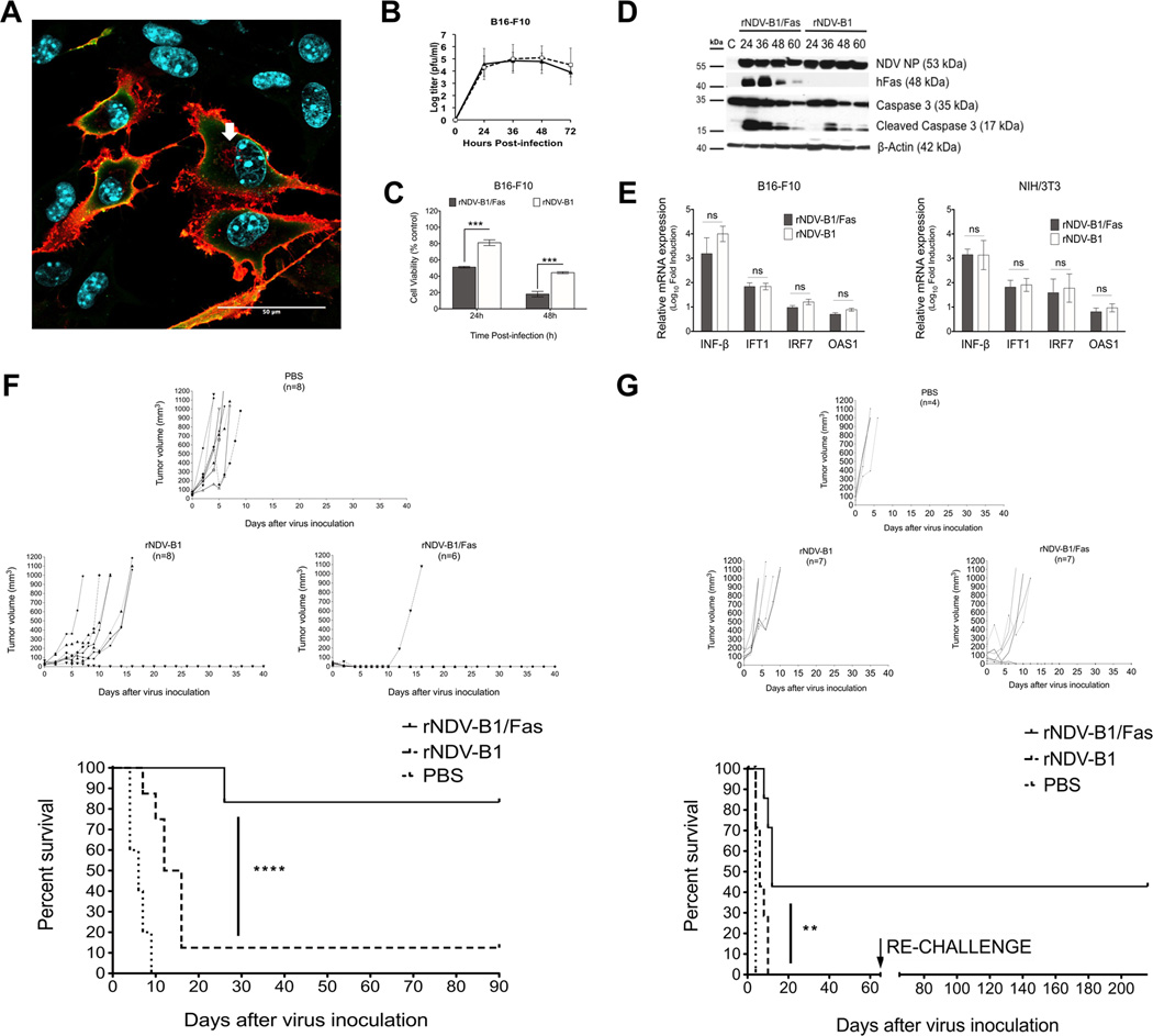
Figure 4. rNDV-B1/Fas virus exerts higher oncolytic capacity and increases survival in a syngeneic melanoma murine model.
A, immunofluorescence detection of human Fas receptor in rNDV-B1/Fas-B16-F10 infected cells. Confocal microscopy image of murine melanoma B16-F10 cells infected with rNDV-B1/Fas. Cells were infected at an MOI of 1 PFU/cell, fixed 20 hours post-infection and stained with monoclonal anti-human Fas antibody (red), polyclonal anti-NDV serum (green) and Hoechst for nuclear contrast. Scale bar 50µm. White arrow points endosome compartment localization of the recombinant human Fas receptor. B, multicycle growth curves. B16-F10 monolayers were infected at an MOI of 0.01 PFU/cell. At different time-points post-infection viral titers in the supernatant were determined. Data points show mean values from three replicates with error bars representing standard deviations. C, cytotoxicity. B16-F10cells were infected at an MOI of 1 PFU/cell and their viability was determined by MTT viability assay at different time points (24, 36 and 48 hours post-infection) (n=3, ***p< 0.0005). D, time course of caspases activation. Western blot. B16-F10 cells were infected with rNDV-B1/Fas and rNDV-B1 at an MOI of 1 PFU/cell and lysates were obtained at different time points post-infection. Apoptosis activation was assessed by western blot using an specific anti-caspase 3 antibody. Human Fas receptor expression was detected using an anti-human Fas monoclonal antibody. Viral replication (NP levels) was detected using an anti-NDV polyclonal serum. E, Interferon response induction in infected cells. Monolayers of B16-F10 and NIH/373 cells were infected with the virus suspension at an MOI of 1 PFU/cell.
Total RNAs from cultured cells were isolated 8 hours post-infection. Mean n-Log10 fold expression levels of cDNA from three individual biological samples, each measured in triplicate, were normalized to 18S rRNA levels and calibrated to mock-treated samples. mRNA expression levels for INF-beta, IFT1, IFR7 and OAS1 were evaluated in both cell lines (ns> 0.05). F, oncolytic capacity of rNDV-B1/Fas and rNDV-B1 viruses in syngeneic murine melanoma tumor model. Tumor growth curves and long term survival report. B16-F10 cells were implanted in the flank of the posterior right leg of C57BL/6 mice. Starting on day ten after tumor cell line injection, the animals were intratumoral treated every other day with a total of three doses of 5×10⁶ PFU of rNDV-B1/Fas, rNDV-B1 or PBS for control mice. Tumor volume was monitored every 48 hours or every 24 hours when approaching the experimental end point of 1000 mm3, after which mice were euthanized (****p< 0.0001). G, long term survival and protection against melanoma re-challenge. Syngeneic melanoma tumors and treatment were performed as described in F. After 30 days of absence of tumor, B16-F10 cells were re-implanted in the flank of the opposite leg. The new development or relapse of tumors was reviewed periodically up to 6 months.

