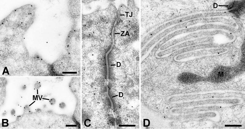
Figure 2.
Electron microscopic localization of ezrin in striated duct cells. Gold particles indicating ezrin reactivity are associated with the luminal membrane (A) and microvilli (MV) (B), the lateral membranes especially in relation to intercellular junctions and associated cytoskeletal components (C), and the basal membrane infoldings (D). Tight junction (TJ); zonula adherens (ZA); desmosome (D); mitochondrion (M). Scale bars = 0.25 μm.

