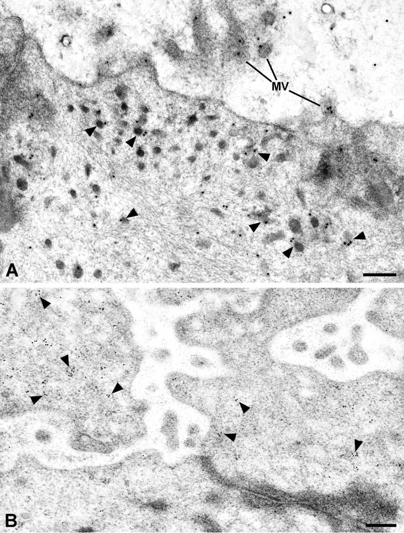
Figure 3.
Electron microscopic localization of PKA in striated duct cells. A: RII subunits of PKA are present along the luminal membrane and microvilli (MV), and are associated with apical granules and vesicles (arrowheads). B: Catalytic (C) subunits of PKA are mainly associated with apical granules and vesicles. Scale bars = 0.25 μm.

