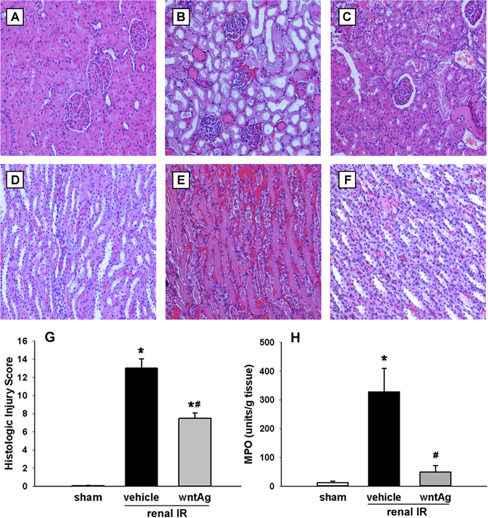
FIG. 3. Effect of Wnt agonist on kidney histologic architecture and MPO-induced damage after renal IR.
Kidneys were harvested 24 h after reperfusion, fixed, and stained with hematoxylin-eosin. Representative photomicrographs at 200× magnification of cortex and medulla, respectively, of (A, D) sham, (B, E) vehicle, and (C, F) Wnt agonist-treated (wntAg) groups. (G) Semi-quantitative histologic injury score measuring differences in dilation or loss of Bowman’s space, flattening of renal tubular epithelium, loss of tubular brush border, microhemorrhage, and tubular casts examined on standard hematoxylin-eosin staining as described in Materials and Methods. (H) Kidney tissue myeloperoxidase (MPO) activity was determined by spectrophotometer. Data presented as means ± SEM (n=4–6/group) and compared by one-way ANOVA and SNK method; *P < 0.05 vs. sham; #P < 0.05 vs. vehicle.

