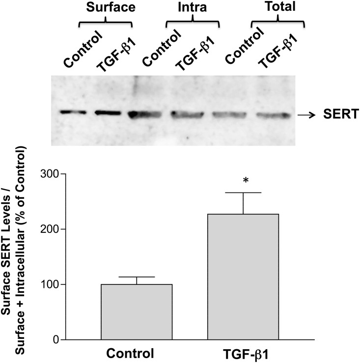
Fig 3.
A. TGF-β1 increases surface expression of SERT in Caco-2 cells. Biotinylated proteins were run on SDS-polyacrylamide gel. The blot was immunostained with anti-SERT antibody. Representative blot of 3 separate experiments is shown. B. Densitometric analysis of SERT surface expression. Results are expressed as surface SERT/ total SERT (Surface + Intracellular). Values represent mean ± SEM of 3 different experiments. *p<0.05 or less compared to control.

