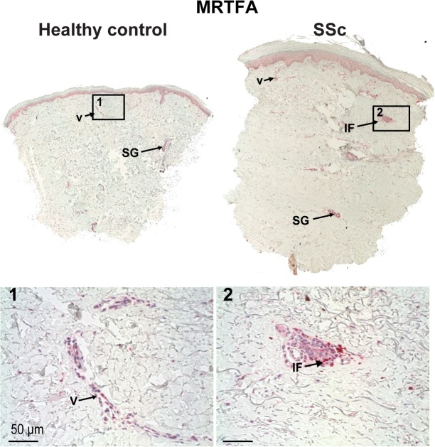
Fig 1. MRTF-A expression is more prominent in SSc skin then healthy control.
MRTF-A staining (1:1000) of healthy control (left) and SSc (right) sections. Several pictures across the wound were merged to produce panoramas of whole sections. Original magnification 10X. Expression of MRTF-A is increased in SSc sections, with more seen in dermal cells, keratinocytes, and vasculature especially within inflammatory foci around small vessels in SSc. Arrow indicate sweat and sebaceous glands (SG), vascularture (V) or inflamatory foci (IF) stained with MRTFs. 1. MRTF-A vascular cell staining in healthy controls or 2. pervascular inflammtory foci in SSc. (30X).

